Home » Without Label » Tendon Diagram / Tendons and Ligaments|Injuries|Recovery|Difference|Function / A tendon is a band of tissue that connects a muscle to a bone.
Tendon Diagram / Tendons and Ligaments|Injuries|Recovery|Difference|Function / A tendon is a band of tissue that connects a muscle to a bone.
Tendon Diagram / Tendons and Ligaments|Injuries|Recovery|Difference|Function / A tendon is a band of tissue that connects a muscle to a bone.. This diagram depicts muscle in the body 744×1054 with parts and labels. The achilles tendon is a tough band of fibrous tissue that connects the calf muscles to the heel bone (calcaneus). You can see a diagram of the achilles tendon below. Bones in shoulder, ligaments of the shoulder joint, parts of the shoulder joint, shoulder anatomy, shoulder joints and muscles, shoulder structure anatomy, shoulder tendon anatomy, shoulder tendons ligaments, human muscles, bones in shoulder, ligaments of the shoulder joint, parts of. Attaches the calf muscles to the calcaneus, most important muscles for running, jumping, walking etc.
This sudden, tight, intense lower leg pain is sometimes called a charley horse. Your biceps tendons attach the biceps muscle to bones in the shoulder and in the elbow. Ligaments join the knee bones and provide stability to the knee: The achilles tendon enables us to walk, without it we would not be able to raise our heels of the ground. This important tendon in the back of the calf and ankle connects the plantaris, gastrocnemius, and soleus muscles to.
Patient Care & Pain Treatment Solutions | Arthritis ... from www.arthritisandinjurycare.com A muscle's origin is where a tendon attaches it to the *less* movable bone. Jul 05, 2018 · the foot diagram has a complex structure made up of bones, ligaments, muscles, and tendons. The bones of the hip include the femur, the ilium, the ischium, and the pubis. Tendons are the connection between bones and muscles. In the back and elsewhere in the body, tendons attach muscles to bones. Foot and ankle musculoskeletal key : Tendons are found throughout the body, from the head and neck all the way down to the feet. Allows the action of raising the foot.
Superficial posterior muscles of the forearm posterior compartment muscles of the forearm.
The achilles tendon is the largest. Ligaments join the knee bones and provide stability to the knee: This diagram depicts muscle in the body 744×1054 with parts and labels. Flexor tendon lacerations are classified into five zones 2, 15, 16. Tendon diagrams and design force vectors. 17 photos of the diagram of shoulder muscles and tendons. Bones in shoulder, ligaments of the shoulder joint, parts of the shoulder joint, shoulder anatomy, shoulder joints and muscles, shoulder structure anatomy, shoulder tendon anatomy, shoulder tendons ligaments, human muscles, bones in shoulder, ligaments of the shoulder joint, parts of. This important tendon in the back of the calf and ankle connects the plantaris, gastrocnemius, and soleus muscles to. For images of the muscle, click on each link under location. A tendon is a band of tissue that connects a muscle to a bone. Browse 318 hand anatomy tendons stock photos and images available, or start a new search to explore more stock photos and images. In the back and elsewhere in the body, tendons attach muscles to bones. Tendons are the connection between bones and muscles.
Foot anatomy diagram, foot joint diagram, foot sprain diagram, foot tendons and ligaments pain, leg tendon diagram. Its muscle belly is in the forearm. Foot and ankle musculoskeletal key : Tendons, located at each end of a muscle, attach muscle to bone. By connecting our rigid bones to our powerful muscles, tendons allow us to move.
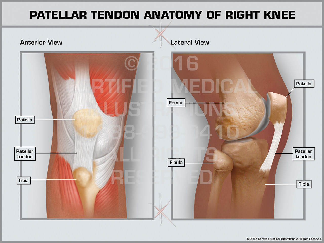
Patellar Tendon Anatomy of Right Knee - Print Quality ... from cdn10.bigcommerce.com This tendon connects the patella (kneecap) to the tibia. Jul 05, 2018 · the foot diagram has a complex structure made up of bones, ligaments, muscles, and tendons. The anterior cruciate ligament prevents the femur from sliding backward on the tibia (or the tibia sliding forward on the femur). Tendons are thick bands of tissue that connect muscles to bones. Tendons attach muscles to bones. The tendon is firmly connected to muscle fibres at one end and to components of the bone at its other end. In the back and elsewhere in the body, tendons attach muscles to bones. These structures work together to support the body, enable a range of movements, and send messages from the brain to.
Allows the action of raising the foot.
There are over two dozen gorgeous and painstakingly detailed illustrations on this chart, from the extensor pollicis longus to the flexor digitorum. For images of the muscle, click on each link under location. Ligaments and tendons are adapted in response to changes in mechanical stiffness. Allows the foot to be turned inward and also supports the arch of the foot. The two peroneal tendons in the foot run side by side behind the outer ankle bone. Allows the action of raising the foot. The hand incorporates countless muscles, bones, tendons and ligaments into simple motion and this chart covers them all. Foot and ankle musculoskeletal key : Intermediate back muscles and c. Bones, cartilage, ligaments, and tendons. The achilles tendon is the strongest and largest tendon in the body. Extends spine and trunk back. Muscles and tendons of the human arm and hand, vintage engraved.
Tendons are found throughout the body, from the head and neck all the way down to the feet. The achilles tendon is also called the calcaneal tendon. On the other hand, the insertion is where a tendon attaches that muscle to the *more* movable bone. Diagram of tendons in forearm. Brings leg back to and across body.
Achilles tendon pain? Which type do you have? - Sundial ... from sundialclinics.co.uk The achilles tendon is the strongest and largest tendon in the body. Movement occurs when our muscles pull on our bones, relocating them. Tendons are the connection between bones and muscles. Tendons are thick bands of tissue that connect muscles to bones. Bones in shoulder, ligaments of the shoulder joint, parts of the shoulder joint, shoulder anatomy, shoulder joints and muscles, shoulder structure anatomy, shoulder tendon anatomy, shoulder tendons ligaments, human muscles, bones in shoulder, ligaments of the shoulder joint, parts of. Your biceps tendons attach the biceps muscle to bones in the shoulder and in the elbow. Medical labeled diagram closeup with muscle, transverse carpal ligament, median nerve, tendon sheath, flextor tendons and bones. Raises heal when leg is straight.
This important tendon in the back of the calf and ankle connects the plantaris, gastrocnemius, and soleus muscles to.
The pubis, ischium, and ilium together constitute the pelvis while the thigh bone is the femur. Allows the foot to be turned inward and also supports the arch of the foot. Brings hip away from body. Diagram of tendons in forearm. These structures work together to support the body, enable a range of movements, and send messages from the brain to. A tendon is a band of tissue that connects a muscle to a bone. Superficial posterior muscles of the forearm posterior compartment muscles of the forearm. Tendon diagrams and design force vectors. The anterior cruciate ligament prevents the femur from sliding backward on the tibia (or the tibia sliding forward on the femur). Foot anatomy diagram, foot joint diagram, foot sprain diagram, foot tendons and ligaments pain, leg tendon diagram, peroneal tendonitis, foot, foot anatomy diagram, foot joint diagram, foot sprain diagram, foot tendons and ligaments pain, leg tendon diagram, peroneal tendonitis. This tendon connects the patella (kneecap) to the tibia. Intermediate back muscles and c. The tendon runs down the back of your lower leg from the back of the knee to the heel.